Sunday, October 10, 2021

Bladder Cancer Glossary Organized by Topic


As you are educating yourself on all things bladder cancer, you will come upon some medical jargon that can be really confusing. This will especially be the case when you are on cancer forums or Facebook groups. Or even this will be the case sometimes when speaking with your doctor or nurse.
 
The list I have made below is grouped into topic sections. When I study, it's helpful to have similar words together. I do list some pretty basic things here, because some people really don't even know the basics. If there are more terms you would like added to this list, leave it in the comments below.   
 
BCAN (Bladder Cancer Advocacy Network) has a great and extensive glossary of terms that is alphabetical, and not sectioned like I have here. Click here to go there.  
 
I hope this list can help you. I know it's a lot. Take a deep breath. Bookmark this page, so you can refer back to it whenever you need. You can do this! 💗 


Different Types of Physicians and Specialists for Bladder Cancer 
🔶 Primary Care Physician / PCP / Primary Care Provider/ GP / General Practitioner: They are healthcare providers or practitioners that see people that have common medical problems. This is the doctor you go to for your yearly check up. A primary care provider can be a physician or Nurse Practitioner, also called an NP. If you are concerned you have symptoms of bladder cancer, you will usually start by seeing your primary care provider. 
🔶 Urologist: A physician who specializes in diseases of the urinary tract system in men and women and in male reproductive organs. They are urinary surgeons. If you are concerned you have symptoms of bladder cancer, you will go to urologist to get an overall urinary assessment, and most likely a cystoscopy/ TURBT/ biopsy and verify that you have cancer. Many urologists treat bladder cancer. However, if your cancer is of a higher stage, you might want to seek out a urologist oncologist. Cystoscopy is a scope with a camera and light that look in your bladder. A TURBT is a surgery where you are put asleep (general anesthesia) and the tumor is taken out. The will also put you to sleep for bladder biopsies, where they will take small tissue samples of your bladder and test for cancer cells. 
🔶 Urologist Oncologist: a urologist who has special training in diagnosing and treating cancers of the male and female urinary tract, and the male reproductive organs.   
🔶 Oncologist / Medical Oncologist: A doctor who specializes in diagnosing and treating cancer using chemotherapy, hormonal therapy, and immunotherapy/biological therapy. A medical oncologist often serves as the main caretaker of someone who has cancer and coordinates treatment provided by other specialists.
🔶 Radiologist: Radiologists are medical doctors that specialize in diagnosing and treating injuries and diseases using medical imaging (radiology) procedures (exams/tests) such as X-rays, computed tomography (CT), magnetic resonance 
imaging (MRI), nuclear medicine, positron emission tomography (PET) and ultrasound.
 🔶 Pathologist: A pathologist is a board certified physician who studies body fluids and tissues, identifying diseases by studying cells and tissues under a microscope. When your tumor or biopsy is taken out, it's sent to a pathologist to look at, and determine if it's cancer or not.
🔶  Radiation Oncologist: A doctor who specializes in using radiation to treat cancer.


Basic Anatomy and Bladder Cancer Terms 
🔶  Acute: it is usually abrupt or a sudden onset or start of a condition or disease. Often there is a lot of pain. It also usually will mean that the illness will be of short duration. 
🔶  Abdomen / Abdominal Cavity: The part of the body that contains the pancreas, stomach, intestines, liver, gallbladder, and other organs. When the bladder is full of urine, it will extend into the abdominal cavity. However, the bladder is located in the region of the pelvis. 
🔶 Anesthesia / General Anesthesia / Local Anesthesia / Regional Anesthesia: this means that you can't feel pain. When used in the medical world, it's usually the pain medication that is given before procedures and surgery. General anesthesia is when the patient is unconscious and feels nothing. The pain medications are given by gas (breathing it) or by IV (intra venous or in your vein). Local anesthesia is when the patient is wide away during surgery. Medicine is injected to numb a small area. regional anesthesia is when the patient is awake, but parts of the body are asleep. For instance, this would be the numbing medicine used on your urethra, when you are getting a cystoscopy, or scope that looks in the bladder. 
🔶  Bladder / Urinary Bladder: A hollow, expandable organ in the pelvic region with flexible, muscular walls. It stores urine before it is excreted, or let out of the body.
 🔶  BC / Bladder Cancer: Cancer that forms in the bladder. There are different kinds, different stages, and different grades. Look below for definition and information about these things.
 🔶 Cancer: A term for diseases in which abnormal cells divide without control. Cancer cells can invade nearby tissues and can spread through the bloodstream and lymphatic system to other parts of the body. You can only be certain a mass or tumor is cancer, by sending a sample of it to pathology. 
🔶 Carcinogen: any substance that causes cancer. 
🔶 Chronic: a medical condition that has been lasting a long time. Usually that means it's lasting 3 months or longer.
🔶 Clinical Trial: A research study that tests how well new medical treatments or other interventions work in people. Each study is designed to test new methods of screening, prevention, diagnosis, or treatment of a disease. 
🔶 Fallopian Tubes: Part of the female reproductive tract. The long slender tubes through which eggs pass from the ovaries to the uterus. Many times when the bladder is removed, these are taken out as well.
🔶 Kidneys: a pair of organs in the abdominal cavity that excrete, or expel urine as waste. They are the beginning of the urinary system. 
 🔶 Lymph Node / Lymph / Lymphatic System / Lymph Gland / Lymph System: A small bean-shaped structure that is part of the body's immune system. Lymph nodes filter substances that travel through the lymphatic fluid, and they contain lymphocytes (white blood cells) that help the body fight infection and disease. There are hundreds of lymph nodes found throughout the body. They are connected to one another by lymph vessels. Clusters of lymph nodes are found in the neck, axilla (underarm), chest, abdomen, and groin. For example, there are about 20-40 lymph nodes in the axilla. The lymphatic system is the tissues and organs that produce, store, and carry white blood cells that fight infections and other diseases. This system includes the bone marrow, spleen, thymus, lymph nodes, and lymphatic vessels (a network of thin tubes that carry lymph and white blood cells). Lymphatic vessels branch, like blood vessels, into all the tissues of the body. Also called lymph system. When aggressive and large cancers are taken out, usually nearby lymph nodes are taken out as well, and they are checked to see if cancer has spread to them. When the bladder has to be removed because of bladder cancer, some lymph nodes are taken and sent to pathology.
 🔶 Ovaries: The pair of female reproductive glands in which the ova, or eggs, are formed. The Ovaries are located in the pelvis, one on each side of the uterus.
🔶 Pelvis / Bony Pelvis / Pelvic Girdle / Pelvic Cavity: The pelvis or bony pelvis or pelvic girdle is a basin-shaped complex of bones that connects the trunk and the legs and supports and balances the trunk. The space between these bones is called the pelvic cavity. The pelvic cavity is a funnel-shaped space surrounded by pelvic bones and it contains organs, such as the urinary bladder, intestines, and rectum (8 inch chamber that connects the colon to the anus). In females, it also contains the vagina, cervix, uterus, fallopian tubes, and ovaries. In males, it also contains the prostate and seminal vesicles.. The pelvic cavity and the abdominal cavity together form the larger abdominopelvic cavity.
🔶 Penis: the male genital organ. It consists mostly of erectile tissue, that allows for elimination of urine, and the transfer of sperm during sex, both through the urethra. The urethra is the hole at the end of the penis. Please see picture above.
🔶 Prostate: a glad surrounding the neck of the bladder in males. It releases prostatic fluid, which is part of the fluid in the semen. It's released during ejaculation of the semen, during sex. 
 🔶 Side Effects: Problems that occur when treatment affects healthy cells. Common side effects of bladder cancer treatment (depending on the treatment) are fatigue, bleeding, hematuria (blood in urine), urinary urgency, urinary frequency, bladder spams, nausea, vomiting, decreased blood cell counts, hair loss, incontinence, inflammation, mouth sores, etc. There is a section below that talks about all of these and many more.
🔶 Trigone of Bladder / Vesical Trigone / Trigone: The smooth, triangular area  inside the bladder where the ureters and urethra connect to the interior of the bladder. It is sensitive to expansion and signals the brain of the need to empty.
🔶 Ureter: the tube that carries urine from the kidney to the bladder. You have two of these, one for each kidney. They attach on the bottom of the kidneys and the top of the bladder.
🔶 Urethra: this is the tube that attaches to the bottom of the bladder. It takes the urine from the bladder and out of your body. This is the hole that you will see when you look at a penis, or when you spread the labia of a woman. This is where catheters go in to access the bladder. This is also where the cystoscope will go in to access the bladder with a camera.
🔶 Urinary Tract: the body's drainage system for removing urine. Each pare of the urinary tract must work together in the correct order. The main organs of the urinary tract include the kidneys, ureters, bladder, and urethra.
🔶 Urine: liquid waste produced by the kidneys. It contains water, salt, urea, and other waste products removed from the blood. Usually it is yellowish in color. Once produced in the kidneys, it travels through the ureters, where it is stored in the bladder and then excreted, or let out of the body, through the urethra.
 🔶 Urothelium / Urothelial (yoo-roh-THEE-lee-um): the lining of the urinary tract is called the urothelium. There are urothelial cells that make up the urothelium. So when you see the terms urothelial carcinoma (cancer tumor) put together, it means that the cancer is in the lining of the urinary system - the urothelium. The lining of the urinary tract includes the renal pelvis (the internal parts of the kidney, see pictures above), ureters, bladder, and urethra. Unfortunately, because bladder cancers include the urothelial layer, you are at a potential risk of developing cancer in the other areas of the urinary system that has an endothelial lining. Meaning the cancer can pop up in the kidneys, ureters, bladder, or urethra at a later time. That is why a CT (cat scan) needs to be done at least yearly to rule out the cancer growing in another area of your urinary system.
🔶 Uterus: the organ in the lower body of a woman, where offspring are conceived and in which they grow. Also called the womb. The uterus is located directly behind the bladder. 
🔶 Vagina: the muscular tube leading from the external genitals to the cervix of the uterus in women. If your bladder has to be removed, many times a part of the vagina is also removed and has to be reconstructed. This can cause concerns and issues surrounding having sex again. 
🔶 Void / Urinate / Pee / Wee: to expel urine from the body. If you get on bladder cancer forums and groups, you will so those from the UK use the term wee. 😀


 Signs and Symptoms of Bladder Cancer and Side Effects of Treatments
 🔶 Acute Urinary Retention: This is an emergency! It's when suddenly you are not able to urinate. Most of the time its because the bladder or urethra cannot let the urine pass. This can be from trauma to the bladder (making in able to contract and press the urine out) or trauma to the urethra (it swells up and clamps shut). A blood clot, sloughing from a wound in your bladder, or piece of tumor can also block the way. This can also happen after you have BCG (it's quite rare). I had an experience with this. Click here to read more.
 🔶  Anemia: a condition that happens when you don't have enough red blood cells or hemoglobin in your blood. In bladder cancer, that would usually mean that is happening because of long term bleeding. This will result in weakness, and pallor or unhealthy pale appearance. This can be a sign of bladder cancer.  
 🔶  Bladder Spasms (medical term is detrusor contraction) / Overactive Bladder: this is when your bladder squeezes or contracts uncontrollably, suddenly without warning, causing an urgent need to release urine. This can cause frequent and urgent need to urinate. The spasm can force urine from the bladder, causing leakage. These can be so intense and painful, it can feel like a uterine contraction (if you are female and have experienced that before), and can last seconds to 10 minutes or more. There are medications that can help with this. This can be a sign of bladder cancer.
 🔶 Chronic Urinary Retention: This is when you can't empty your bladder all the way, but you can usually still get some urine through. It's very uncomfortable, and it's persistent. This can be a sign of having bladder cancer.  
 🔶 Continence / Incontinence: Continence is the ability to control the flow of urine from the bladder to the outside of the body. You can usually control urination due to muscular structures called sphincters, which wrap around the base of the bladder and urethra. Additionally, if the bladder is damaged, then the bladder cannot contract properly to help expel the urine from the body. Removal of or damage to the urinary sphincters or the bladder, can result in an inability to control the flow of urine normally. That is what incontinence is. It's when you don't have control of urinating, and you wet yourself.
🔶 Cystitis: cystis means bladder, and itis means inflammation. So cystitis means inflammation of the bladder. BCG, UTI's, chemo, surgery and other bladder cancer treatments can cause this. Severe cystitis can make it so the bladder won't contract and push urine out any longer, meaning you can't urinate. If this is ever the case, it's an emergency, and you need immediate care. Chronic, or long term cystitis might be a sign of bladder cancer. 
🔶 Flank Pain / Lower Back Pain: this is the pain in the lower back and/or side. The pain is most likely coming from your kidneys. This can be a sign of bladder cancer.  
🔶 Gross Hematuria / Microscopic Hematuria: Heme is blood, and uria is referring to your urine. So hematuria means blood is in your urine. This is the most common symptom of bladder cancer. When looking at your urine sample, they find red blood cells in your urine. Gross hematuria means that when anyone looks at the urine sample, you can see blood. Microscopic hematuria means that under a microscope you can see blood cells in your urine, but the blood can't be visualized with a human eye. The color can range from slight pink, to bright red, to brownish (older oxidized blood), to orange. 
🔶  Hair Loss / Hair Thinning / Brittle and Dry Hair: Depending on the chemotherapy, you can loose all your hair.  Some people have reported that BCG can cause hair thinning and brittle or dry hair. Increase your protein, take collagen everyday, and invest in a good hair oil to help protect and moisturize your hair. 
🔶  Permanent Hearing Loss: this is a potential side effect of chemotherapy.
🔶  Impotent: a male that is unable to have an erection adequate for sexual intercourse. Sometimes this can be a side effect of removing the bladder or prostate.
🔶  Inflammation: a reaction that can cause symptoms such as swelling, soreness, redness, and warmth often as a reaction to illness or injury. BCG, UTI's, chemo, surgery and other bladder cancer treatments can cause this.
🔶  Kidney Stone / Renal Calculus: a hard mass formed in the kidneys, typically consisting of calcium compounds that can't be broken down by the body. Usually kidney stones have to be "passed" meaning they need to come out through your urinary system... kidney, ureter, bladder, then urethra. They are usually very painful. Sometimes, people might think what they passed was a kidney stone, but it might be a piece of a tumor in your urinary system.
🔶 Nerve Pain / Neuropathy / Neuropathic Pain : disease or dysfunction of one or more peripheral nerves, typically causing numbness or weakness. Chemo can cause this permanent dysfunction. 
🔶 Overactive Bladder: When your bladder spasms very often, causing someone to need to urinate all the time. There can also be urine leakage associated with this as well. There are medications that can help with this. This can be a sign of bladder cancer.
🔶 Urethritis: your urethra is inflamed (itis means inflammation). If it gets severe enough, it can clamp shut, thus blocking the flow of urine. If this happens, it's an emergency. It's called acute urinary retention.
🔶 Urge Incontinence: when your bladder spasms, and you leak urine because of the urgent need to urinate. There are medications that can help with this.
🔶 Urinary Frequency and Urgency: it's exactly what it sounds like. You have an incredibly strong urge to urinate very often. These symptoms will come after a TURBT surgery (take out the tumor by going up your urethra - OUCH!), after having a catheter for treatments or tests, or if you have a urinary tract infection. This can also be a possible sign of bladder cancer, especially if after taking antibiotics the pain doesn't go away.
🔶 Urinary Tract Infection / UTI: it's an infection in any part of your urinary system - kidneys, ureters, bladder, and urethra.


Tests Associated with Bladder Cancer

🔶 CT / Cat Scan / Computerized Tomography : CAT scan is what most people call it.  A computerized tomography (CT) scan combines hundreds of X-ray images taken from different angles around your body and uses computer processing to create cross-sectional images (slices) of the bones, blood vessels and soft tissues inside your body. CT scan images provide more-detailed information than plain X-rays do. When you see a CT machine, you will see a large machine that has a bed going through a small tube. When you get the scan, you will lie down on the bed, and it moves back and forth.
🔶 CT With and Without Contrast: CT scans may be done with or without "contrast." Contrast refers to a substance taken by mouth or injected into an intravenous (IV) line that causes the particular organ or tissue under study to be seen more clearly, or looks like it lights up when looking at the X-rays from the CT. Having a CT scan with contrast will sometimes allow a 3D image to be created. IV contrast, often an iodine contrast solution, can be very hard on your kidneys. Be sure to have your kidney function tested before getting a CT scan. 
🔶 CT Urogram : CT scan specifically for the urinary system. This is often done with contract and without contrast.  
🔶 Cystography (sist-o-gra-fee) / Cystourethrography (sist-o-ur-eth-rah-gra-fee) / Voiding Cystourethrography / CT Cystography: This test is more unusual for bladder cancer, but might be used for other urinary issues related to bladder cancer and treatments. Cystography is an imaging test that can help diagnose problems in your bladder. It uses X-rays. They may be X-ray pictures or fluoroscopy, a kind of X-ray "movie." During cystography, the healthcare provider will insert a thin tube called a urinary catheter that goes into your urethra, and inject contrast dye into your bladder. The contrast dye will let the healthcare provider see your bladder more clearly. He or she will take X-rays of the bladder. Cystography is sometimes combined with other procedures. For example, cystourethrography images the bladder and the urethra. The healthcare provider may also use fluoroscopy to watch how the bladder empties while you urinate (voiding cystourethrography). Cystography may show whether any urine backs up into the kidneys (vesicoureteral reflux). CT (cat scan) cystography is sometimes used following trauma or recent surgery.
🔶 Cystoscopy (sis-TOS-kuh-pee) / Scope / Cysto / Cystoscope / Rigid Cystoscope / Flexible Cystoscope:  A cystoscopy is a procedure that allows your doctor to examine the lining of your bladder and the urethra (the tube that carries urine out of your body). Some people will call it for short a scope or a cysto. A hollow plastic tube, called a cystoscope is equipped with a camera and light, and is inserted into your urethra and slowly advanced into your bladder. This is the gold standard test for ruling out bladder cancer. This can be done in the doctor's office with numbing gel, using a flexible cystoscope, which is much smaller than the one they use for surgery. Under general anesthesia (when you are put to sleep during surgery) they use a rigid cystoscope, which is much larger and painful, thus the need for general anesthesia.  If you are getting treatment for bladder cancer, buckle up and get used to these. For the first two years, you will get them every 3 months, then 6 months, then yearly. 
🔶 Imaging : Tests that produce pictures of areas inside the body.
🔶 MRI / Magnetic Resonance Imaging: MRI's will also be used in diagnosing and treating bladder cancer. A MRI is a type of scan that uses strong magnetic fields and radio waves to produce detailed images of the inside of your body. The machine contains powerful magnets, so you will not be allowed to wear any metal when getting a scan. MRI's are non-invasive. When you see a MRI machine, you will see a large machine that has a bed going through a long tube. When you get the scan, you will lie down on the bed, and it moves inside the long tube. Once in position, you will stay in that spot for quite a long time, being asked not to move generally. That can be from 10 minutes to 30 minutes. During that time you will hear a series of repetitive sounds. MRI's are especially good tests for soft tissues. 
🔶 PET Scan / Positron Emission Tomography : uses tracers (radioactive drug) to show normal and abnormal metabolic activity. That's a fancy way of saying that it will find really fast growing cells - like cancer. When looking at the images, these cells would light up. It can also help identify heart disease and brain disorders. PET scans must be interpreted carefully because noncancerous conditions can look like cancer, and some cancers do not appear on PET scans.
🔶 Utrasound / Sonogram / Ultrasonogram : A computer picture of areas inside the body created by bouncing sound waves off organs and other tissues. It can be useful in determining the size of a bladder cancer and whether it has spread beyond the bladder to nearby organs or tissues. It can also be used to look at the kidneys. This is usually an easy test to have, and it uses no radiation.
🔶 Pyelogram (PIE-uh-low-gram) / Intravenous Pyelogram / IVP / Excretory Urogram: an intravenous pyelogram, is an X-ray exam of your urinary tract. These are often used to diagnose kidney stones, bladder stones, enlarged prostate, kidney cysts, or urinary tract tumors. During this exam you will have X-ray dye (iodine contrast solution) injected into a vein in your arm.
For an intravenous pyelogram, you lie on your back on an exam table. The X-ray machine usually is either attached to or part of the table. An X-ray image intensifier — the part of the machine that obtains the images — is positioned over your abdomen. X-rays are taken before the contrast, and then the dye is injected and more images are taken at specifically timed intervals. Toward the end of the exam you may be asked to go to the bathroom and urinate. Then you return to the exam table, so that the health care team can get an X-ray of your empty bladder.
🔶  Urinalysis (Ur-i-nal-i-sis) / UA / Urine Dip / Urine Dipstick Test: Urinalysis is a urine sample that is looked at under a microscope. Sometimes this sample can also also be taken in the doctors office, and that is called a urine dipstick test. They will use small testing strips that can tell them quickly information about your urine including if there is any blood in the sample, or if there are white blood cells in the sample (meaning you have an infection in your urinary tract, or UTI). The sample can be taken with just washing off your genitals before urinating in a cup. It can also be taken when there is a catheter or scope inside your bladder. 
🔶 Urine Cytology : Urine cytology is a test to look for abnormal cells in your urine. It's used with other tests and procedures to diagnose urinary tract cancers, most often bladder cancer. Your doctor might recommend a urine cytology test if you have blood in your urine (hematuria). These tests will often be ordered when you get a cystoscopy. 

Pain Medications to Help With Bladder Cancer

🔶 Acetaminophen / Tylenol 1000mg every 8 hours: over the counter, and helps with pain, muscle aches, and fevers. Don't ever take more than 4 grams in 24 hours. This can be taken every 8 hours. This is also often in narcotics. Make sure you are adding up the TOTAL Tylenol usage for the day. Tylenol is processed in your liver, so can be taken at the same time as Ibuprofen. 

🔶 Ibuprofen / Advil / Motrin 600 mg every 6-8 hours:  over the counter, and helps with pain, muscle aches, inflammation, and fevers. This can be taken every 6-8 hours. Ibuprofen is processed in the kidneys, thus it's okay to take Tylenol at the same time (Tylenol is process in the liver). Make sure you are drinking enough water to not stress your kidneys. Ibuprofen can cause irritation and bleeding in your stomach. This is classified as an NSAID (non-steroidal anti-inflammatory drug). DO NOT COMBINE WITH ASPIRIN!

🔶 Aspirin / Acetylsalicylic Acid / Bayer / 500mg every 6 hours : over the counter, and helps with pain, muscle aches, inflammation, and fevers. Aspirin is processed in the kidneys, thus it's okay to take Tylenol at the same time (Tylenol is process in the liver). Make sure you are drinking enough water to not stress your kidneys. Aspirin can cause irritation and bleeding in your stomach. This is classified as an NSAID (non-steroidal anti-inflammatory drug). DO NOT COMBINE WITH IBUPROFEN!

🔶 Narcotics / Hydrocodone (Vicodin, Norco, Lortab) / Oxycodone (Percocet, Percodan): These are all serious pain killers. You will get them after surgery if you need them. I found that I needed on pill prescribed for BCG #12. It's about a 4-6 hour window that the pain is just intolerable.

🔶 AZO / Pyridium / Phenazopyridine / Prodium / Pyridate / Baridium / Uricalm / Urodine / UTI Relief: a dye that works as a painkiller to sooth the lining of the urinary tract. It helps relieve pain, urgency, and frequency when urinating. It will turn your urine orange while it's in your system. This is totally normal. 😊 It does stain underwear permanently, so get some undies you can throw away. You can get this over the counter. If you want a pill with a higher dose (instead of taking many of the over the count types) you can have your doctor order it for you. 

🔶 Anti-Anxiety : There can develop some real anxiety around tests, procedures and treatments. Lots of these things are painful, and so we develop anxiety about them, often making the pain even worse. So take that anxiety medication! Let someone else drive you to the appointment. 💗


Overactive Bladder or Bladder Spasm Medications

These are medications that help with bladder spasms or overactive bladder. This can help after BCG therapy and after a TURBT. The ER or XL stands for extended release. It makes it so you take the medication once a day, and it release  that medicine throughout the day.

🔶 Toterodine / Detrol 

🔶 Oxybutynin (oxy-BYOO-ti-nin) / Oxybutynin ER / Ditropan XL

🔶 Trospium / Sanctura

🔶  Solifenacin / Vesicare

🔶 Darifenacin / Enablex

🔶 Fesoterodine / Toviaz

🔶 Mirabegron / Myrbetriq


Common Jargon, Types, Stages, and Grades of Bladder Cancer

🔶 Benign Tumor / Benign / Benign Cancer : a non-cancerous tumor. Benign tumors may grow large but do not spread into, or invade, nearby tissues or other parts of the body.

🔶 Cancer: A term for diseases in which abnormal cells divide without control. Cancer cells can invade nearby tissues and can spread through the bloodstream and lymphatic system to other parts of the body. You can only be certain a mass or tumor is cancer, by sending a sample of it to pathology. 

🔶 Cancer Grade / Grade / Grade 1 = Low Grade / Grade 2 = Moderate Grade / Grade 3 = High Grade : there are three cancer grades. Grading is about how the cancer cells look under a microscope compared with normal cells. The more irregular the cells, the higher the grade. The higher the grade, the more aggressive the cancer is. The grade also tells how quickly the tumor is likely to grow and spread. The higher the grade usually the faster the tumor will grow and spread, and the more likely that the cancer will come back (recurrence) and progress (into a higher stage or metastasize forming other cancers in the body). Grading systems are different for each type of cancer.

 🔶 Cancer Stage / Stage / Staging : The stage is determined by the cancer growth in the bladder wall and how far it has spread to nearby tissues and other organs, such as the lungs, the liver, or the bones. Staging is performing exams and tests to learn the extent of the cancer within the  body, especially whether the disease has spread from the original site to other parts of the body.

🔶 Carcinogen: Any substance that causes cancer. 

🔶 Carcinoma : Cancer that forms in the skin or lining of internal organs. In bladder cancer the lining of the bladder is called the urothelium.

 🔶 Clean Margins: this means that when a tumor is taken out, at the outer edge of the tissue removed, there are no cancer cells found. If you have clean margins that will mean that that got it all!  

🔶 Gene / DNA:  Genes are the functional and physical unit of heredity passed from parent to offspring. Genes are pieces of DNA, and most genes contain the information for making a specific protein. 

 🔶 Malignant Tumor: a cancerous tumor. Malignant tumors can spread into, or invade, nearby tissues. They can also spread to other parts of the body through the blood and lymph systems. Also called neoplasm.

🔶 Metastasize / Mets / Metastasis / Metastases / Secondary Tumor / Primary Tumor: Metastasize means to spread form one part of the body to another. When cancer cells metastasize they form what is called a secondary tumor. The cells in the metastatic tumor are like those in the original (primary) tumor. Metastasis is the spread of cancer from one part of the body to another. The plural is metastases. Often with bladder cancer, if metastases occur, it will be in the liver, lungs, and bones. 

🔶 Muscle-Invasive Bladder Cancer / Invasive Bladder Cancer / Infiltrating Cancer / Invasive Cancer: is when the cancer has grown into deeper layers of the bladder wall (muscle), and beyond, goring into surrounding and healthy tissues. These are more likely to spread to other organs and are harder to treat. This includes stage 2, 3, and 4, with being stage 4 extending past the bladder wall. These tumors have characteristically bad biological behavior and are capable of spreading to other parts of the body without much warning. Accordingly, physicians are constantly on the lookout for evidence of disease spread in patients with invasive bladder carcinomas (cancers in the endothelial lining of the bladder). Invasive cancers are less common than superficial ones, but they unfortunately spread to other parts of the body in about half of the patients who have this invasive disease. 

🔶 Non-Invasive Bladder Cancer / Non-Muscle Invasive Bladder Cancer (NMIBC)/ Transitional Cell Carcinoma (TCC) / Urothelial (yer-o-THEE-ul) Carcinoma: I know, I know... that is whole lot of names! I wish they could simplify things. But when you see one of these named, it really means all of them. This is the cancer found in the tissue that lines the inner surface of the bladder called the urothelium.The bladder muscle is not involved, it has not grown into the muscle layer. That means it is stage 0 or stage 1. This is what about 90 percent of all bladder cancers are.  

🔶 p53 Gene / p53 Tumor Suppressor Gene : This is a gene that can help prevent the development of tumors. Because p53 is essential for regulating DNA repair and cell division, it has been nicknamed the "guardian of the genome." In bladder cancer, alterations or this gene are common, occurring in about 50% of transitional cell carcinoma, also called non-invasive/ non-muscle invasive bladder cancer (NMIBC) or urothelial carcinoma. This is the most common form of bladder cancer (about 90%).  

🔶  Pathology Report / Histology Report / Biopsy Report / Pathology / Pathologist / Histology / Histopathology / Patho / Histo: pathology is the study of disease, and histology is the study of microscopic structure of tissues. Histopathology is the study of diseased cells and tissues using a microscope. It is the bridge between science and medicine. A pathologist is a board certified physician who studies body fluids and tissues, identifying diseases by studying cells and tissues under a microscope. After you get a biopsy (a sample of tissue or tumor) or a tumor is resected (taken out), then that tissue or tumor is sent to pathology. The pathologist will look at it under a microscope and tell you if the growth is benign (non-cancerous) or malignant (cancerous). If it is cancer the pathology report/histology report/biopsy report will tell you the type of cancer, the stage of the cancer, and the grade of the cancer. The pathology report will also tell you that the surgeon had clean margins in the tissue sample. You will often see people just use the word patho or histo when describing these results. Example: "The patho showed NED" NED is no evidence of disease by the way. 😊

🔶 Pre-Cancerous : A group of cells, tissue, mass, or tumor that may turn cancerous if left in place or untreated.

🔶 Primary Tumor: The original tumor or cancer in your bladder. 

🔶 Prognosis: What your chance of recovery is. 

🔶 Recurrent / Recurrence / Recur: To occur again. Recurrence is the return of cancer, at the same site as the original (primary) tumor in another location, after the tumor had disappeared. Recurrent UTI's mean you have a lot of urinary tract infections. More than what might be counted as "normal." Some studies have said that possible recurrent UTI's might be attributed to bladder cancer.

🔶 Refractory: In medicine, describes a disease or condition that does not respond to treatment.

🔶 Remission / Partial Remission / Complete Remission / NED / No Evidence of Disease: remission is that signs and symptoms of your cancer are reduced or gone. Remission can be partial or complete. In partial remission, some, but not all signs and symptoms of cancer have disappeared. This could mean the tumor is smaller. In complete remission, all signs and symptoms of cancer have disappeared, although cancer still may be in the body. NED stands for no evidence of disease. It means the patient is in complete remission.

🔶 Risk Factor: A habit, trait, condition, or genetic alteration that increases a person’s chance of developing a disease.

🔶 Secondary Tumor: A term used to describe cancer that has spread (metastasized) from the place where it first started to another part of the body. Secondary tumors are the same type of cancer as the original (primary) cancer.

🔶 Squamous Cell Carcinoma / Adenocarcinoma / Small Cell Carcinoma/ Sarcoma: these are all very rare types of bladder cancers. Squamous cell carcinomas originates from flat-shaped squamous cells found in the tissue lining of the bladder. Adenocarcinomas originate from mucus-secreting gland cells lining the bladder. Small cell carcinoma occurs in less than 1% of all bladder cancers and is very aggressive and doesn't respond well to treatment, often metastasizing or spreading to other body parts. It come from nerve-like neuroendocrine cells in the bladder. Sarcomas come from the bladder’s muscle cells. 

 🔶 Ta / Stage Ta / Ta0 / Superficial Bladder Cancer: This tells us how the tumor looks and that it is stage 0 (caught very early). It's a non-invasive (not in the muscle) papillary (it sticks out and looks like cauliflower or coral) carcinoma. This type of cancer has grown toward the hollow center of the bladder but has not grown into the connective tissue or muscle of the bladder wall. It is only in the urothelium layer (part that touches the urine) of the bladder. It has not spread to nearby lymph nodes or distant sites. This can easily be removed by a TURBT (a surgery where they go in through your urethra, and take out the tumor). The majority of bladder cancers are superficial, though these progress in only a minority of patients, they do usually recur.

🔶 Tis/ CIS/ Stage Tis / Tis / Carcinoma in Situ / Superficial Bladder Cancer: this tells us how the tumor looks and that it is stage 0 (caught very early). This tumor is flat. It's a non-invasive (not in the muscle) flat carcinoma. These cells are only in the urothelium layer (part that touches the urine) of the bladder. These types of tumors have a aggressive being higher grade, increasing the risk of recurrence (coming back) and progression (getting bigger and growing outside the bladder and spreading to other body parts). The majority of bladder cancers are superficial, though these progress in only a minority of patients, they do usually recur.

🔶 Tumor/ T / Mass / Neoplasm / Neoplasia : an abnormal mass of tissue that forms when cells grown and divide more than they should or do not die when they should. Tumors may be benign (not cancer) or malignant (cancer)

🔶 Upper Tract Urothelial Carcinoma / UTUC: While the majority of bladder cancers (approximately 90-95%) arise in the bladder, the urothelial cells that line the bladder are found in other locations in the urinary system. Sometimes these urothelial cancers can occur in the lining of the kidney or in the ureter that connects the kidney to the bladder. This is known as upper tract urothelial cancer (UTUC) correspond to a subset of urothelial cancers that arise in the urothelial cells in the lining of the kidney (called the renal pelvis) or the ureter (the long, thin tube that connects that kidney to the bladder).


Bladder Cancer Treatments, Surgeries, and Therapies

🔶 TUR / Transurethral Resection : Trans = by way of; urethral = urethra; This is usually done under general anesthesia (they put you to sleep for the surgery). A rigid cystoscope is used, along with resectoscope (see definitions below).

🔶 TURBT / Transurethral Resection of Bladder Tumor: Trans = by way of; urethral = urethra; resection = to take out; bladder tumor = a mass or tumor found in the bladder. It's a surgery in which they remove a tumor in the bladder by going through the urethra. This is usually done under general anesthesia (they put you to sleep for the surgery). A rigid cystoscope is used, along with resectoscope (see definitions below).

🔶 Cystoscopy (sis-TOS-kuh-pee) / Scope / Cysto / Cystoscope / Rigid Cystoscope / Flexible Cystoscope:  A cystoscopy is a procedure that allows your doctor to examine the lining of your bladder and the urethra (the tube that carries urine out of your body). Some people will call it for short a scope or a cysto. A hollow plastic tube, called a cystoscope is equipped with a camera and light, and is inserted into your urethra and slowly advanced into your bladder. This is the gold standard test for ruling out bladder cancer. This can be done in the doctor's office with numbing gel, using a flexible cystoscope, which is much smaller than the one they use for surgery. Under general anesthesia (when you are put to sleep during surgery) they use a rigid cystoscope, which is much larger and painful, thus the need for general anesthesia.  If you are getting treatment for bladder cancer, buckle up and get used to these. For the first two years, you will get them every 3 months, then 6 months, then yearly. 

🔶 Resectoscope:  A flexible instrument that can be inserted through the cystoscope sheath, or tube. The resectoscope has a wire loop that is used to remove the tumor. It also has a tool that uses an electric current to cut, remove, or destroy tissue and control bleeding. This is called fulguration.

🔶 Blue Light Cystoscopy:  It's a normal cystoscopy, but a special blue light on the end of the cystoscope. This is a technology to detect and diagnose tumors in the bladder. It uses a contrast solution that is absorbed by bladder cancer cells and makes them turn bright pink or red under a special blue light. This can be used during a TURBT surgery, to find cancer cells that you human eye can't pick up with just white light that is normally used with a cystoscopy. Some doctors offices are also offering a blue light cystoscopy in their offices. Many are not because of the increased expense for them. Many insurance companies refuse to pay extra for a blue light cystoscopy. 

🔶 Narrow Band Imagine Cystoscopy / NBI :  A new technology that utilizes light of specific blue and green wavelengths. It's thought to improve the physician’s ability to detect small, non-muscle-invasive tumors that may not be seen with conventional cystoscopy. The technology takes advantage of the benefits of varying wavelengths of white light without the need for dyes or drugs. This can be done in the urologists office, or during a TURBT surgery. 

 🔶 Fulguration: Destroying tissue using an electric current. This can be done during a cystoscopy, or during a TURBT to destroy the bladder cancer.

🔶 Perforate / Perf / Perforation: Perforation isa  hole that develops thourgh the wall of a body organ like the bladder. This can happen in surgery. If the bladder is perforated, then a foley catheter is placed for several days, while the bladder whall heals.

🔶 Resection / Excision: surgery to remove tissue or part or all of an organ

🔶 Incision: A cut made in the body during surgery.

🔶 Biopsy / Incisional Biopsy / Core Biopsy / Excisional Biopsy / Needle Biopsy / Fine-Needle Aspiration: The removal of cells or tissues for examination under a microscope. The tissues are sent to pathology, where a pathologist will look at them. When only a sample of tissue is removed, the procedure is called an incisional biopsy or core biopsy. When an entire tumor or lesion is removed, the procedure is called an excisional biopsy. When a sample of tissue or fluid is removed with a needle, the procedure is called a needle biopsy or fine-needle aspiration.

🔶 BCG / Bacillus Calmette Guerin / Tice BCG:  BCG is an active form of tuberculosis. Yes, you read that right. Tice BCG is a specific strain that is used in bladder cancer treatment. In the 1970's some scientists noticed that mice that had active tuberculosis, did not get cancer. Thus was born immunotherapy, a treatment that helps your immune system fight the cancer. BCG treatment is given to those with early-stage bladder cancer, stage 0 and stage 1. Those stages mean the cancer has not got into the muscle, and so it's called non-muscle invasive bladder cancer (NMIBC). BCG is instilled or inserted directly into the bladder, through a catheter (small flexible tube). BCG is the gold standard treatment used to treat early-stage bladder cancer. It is usually given once per week. 

🔶 Immunotherapy / Cancer Immunotherapy / Biological Therapy / Biotherapy / Biological Response Therapy (BRM) : Treatment to stimulate or restore the ability of a person's immune system to fight infection and disease. Also used to lessen side effects that may be caused by some cancer treatments. There are many types of immunotherapy used with different treatments of bladder cancer. The most common type is BCG.

🔶 Catheter / Straight Cath / Self-Cath / Foley Catheter: a catheter is a narrow, flexible tube taht can be used to drain and collect urine from the bladder. It can also be used to infuse medication or fluid into the bladder. A regular catheter or straight catheter or the catheter used for self catheterization, is for a one time use only. A foley catheter has a balloon at the end of it. After the catheter is threaded into the urethra and then into the bladder, the ballon is inflated with saline. This catheter will stay in place for as long as needed. It does need to be periodically changed. The ballon stays inflated until the saline is taken out. The balloon keeps the catheter inside the bladder. A foley catheter might be used for many instances including after surgery if the bladder was perforated (a hole made in the wall), or if you can't urinate after BCG. 

🔶 Maintenance BCG: The schedule of BCG includes an initial 6 weeks once per week, then 3 weeks (once per week) at 3 months, 6 months, 12 months, 18 months, 24 months, 30 months, and 36 months. All the doses of 3 weeks, are considered maintenance BCG doses. 

🔶 Induction BCG: this the initial 6 weeks of BCG that are given after the TURBT. The BCG is given once per week. 

🔶 Intravesical / Intravesical BCG / Intravesical Chemotherapy / Local Therapy: Local therapy is treatment that affects cells in the tumor and the area close to it. Intravesical therapy is a local therapy for bladder cacner. With intravesical therapy, the doctor puts a liquid drug right into your bladder rather than giving it by mouth or injecting it into your blood. The drug is put in through a soft catheter that's put into your bladder through your urethra. The drug stays in your bladder for up to 2 hours. This way, the drug can affect the cells lining the inside of your bladder without having major effects on other parts of your body. BCG, and chemotherapy are common medications used for intravesical therapy to treat bladder cancer.

🔶 Local Thearpy: Local therapy is treatment that affects cells in the tumor and the area close to it.
🔶 Systemic Thearpy: Treatment that uses substances that travel through the bloodstream, reaching and affecting cells all over the body.

🔶 Radiation / Radiotherapy / External Radiation / External-Beam Radiation: A cancer treatment that uses a machine to aim high-energy, high doeses of radiation rays at the cancer. Radiation therapy kills cancer cells, slows their growth by damaging their DNA, or will shrink cancer tumors. 

🔶 Chemotherapy / Chemo: used to treat cancer as anti-cancer drugs. Often is cytotoxic (means that it damages or kills cells). Chemotherapy often is targeted for fast growing cells in your body. It can be given intravenously (IV, or in your vein), or intravesical (put directly into your bladder). That would include cancer, but unfortunately will also target your gut (thus the nausea and vomiting), as well as hair follicles, or the structures in the skin from which hair grows. Hair follicles are some of the fastest growing cells in the body. 

🔶 Intravenous / IV : a therapy in which medicaitons or fluids are given straight into your vein. A device is put in your vein by inserting a needle into your skin, and peircing the vein. The device will be taped and a dressing put over it. This device is called an IV. How the medication is given is also called IV or intravenous. For instance you will see written down Normal Saline IV. That means that they normal saline will be given intravenously.

🔶 Neoadjuvant / Neoadjuvant Therapy: Treatment given as a first step to shrink a tumor bofore the main treatment, which is usually surgery. Examples of neoadjuvant therapy include chemotherapy, radiation therapy, and hormone therapy. It is a type of induction (first or starting) therapy.

🔶 Adjuvant / Adjuvant Therapy: Additional cancer treatment given after the primary, or initial, treatment to lower the risk that the cancer will come back. Adjuvant therapy may include chemotherapy, radiation therapy, hormone therapy, targeted therapy, or biological/immunotherapy therapy.


 Bladder Removal and Urinary Reconstruction Surgery Terms

🔶 Cystectomy / Complete Radical Cystectomy (RC) / Complete RC / Partial RC / Segmental Cystectomy: Radical means total or complete. Cystis means bladder, and ectomy means removal. So a complete radical cystectomy is the total surgical removal of the bladder. When a radical cystectomy is performed, your surgeon needs to reconstruct the urinary tract in one of three ways so that your urine can be eliminated from your body: illeal conduit, continent cutaneous diversion/pouch, and neobladder. Partial cystectomy or segmental cystectomy (they are the same thing) is pretty uncommon, in which with the tumor, only a part of the bladder would be removed. If you want a more detailed and excellent description of this process and options available please visit this website from Bladder Cancer Advocacy Network (BCAN). 

🔶 Urinary Diversion / Incontinent Urinary Diversion / Urinary Reconstruction / Continent Urinary Diversion: Urinary diversions are surgical procedures that create new ways for urine to exit your body. These need to be done because the bladder and other structures are removed. There are three surgeries that do this. They are called illeal conduit, continent cutaneous diversion/pouch, and neobladder. 

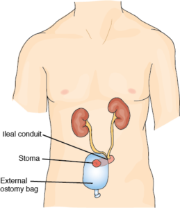
🔶 Ileal Conduit (IC): This is the easiest and most common reconstruction. Your surgeon disconnects a short portion of your small intestine called the ileum. One side of the piece of ileum is attached to a skin opening on the right side of the abdomen and a small stoma, or mouth, is created. A conduit or a passageway between the kidneys and ureters and the urethra is made using the piece of the intestine - the ileum. The ureters (tubes that carry urine from the kidney to the bladder) are attached to one end of the ileum, while the other end of the ileum is attached to the stoma. The stoma is usually located close to the belly button on the right side. A plastic appliance called an ostomy bag, is placed over the stoma to collect urine. Because the nerves and the blood supply are preserved, the conduit is able to propel the urine into the appliance, or ostomy bag.

🔶 Ostomy / Ostomy Bag / Stoma / Urostomy / Ostomate: A stoma is an opening or pathway that your surgeon makes, from the inside of your body to the skin on the outside of your body. A urostomy is a stoma made for your urinary system - so you can get urine out of your body when your bladder has been surgically removed. Your urine will drain into a bag, or ostomy bag, that you put around your stoma. You’ll drain the bag as needed. This is created and used in an Illeal Conduit surgery. There is also a stoma with the continent cutaneous diversion surgery, but there is no bag on the outside to hold the urine. There is a pouch made inside the body that a catheter will access to get the urine out. Someone who has a stoma is called an ostomate. 

🔶 Wound Ostomy Continence Nurse / WOCN Nurse / WOC Nurse / Enterostomal Therapist: a registered nurse that is specially trained to take care of ostomies, and to teach their patients how to manage them is called a wound ostomy continence nurse or WOCN nurse or WOC nurse. They might also help the surgeon figure out the best location for your stoma. An enterostomal therapist is a health professional trained in the care of persons with urostomies and other stomas.

🔶 RC/IC ; Radical Cystectomy With Ileal Conduit: This is the most common urinary system reconstruction. This is the removal of the bladder, with an illeal conduit reconstruction of the urinary tract. You will often see people write RC/IC. 

 🔶 Continent Cutaneous Diversion / Continent Cutaneous Pouch (CCP) / Indiana Pouch Reservoir / Indiana Pouch: This is another method of reconstructing your urinary system, after bladder removal or radical cystectomy. Your surgeon reconstructs a continent reservoir or a urine "storage container," sphere or pouch, using part of your small and large intestine. The ureters are attached to one end of the reservoir, and the other end of the reservoir is connected to a small opening (stoma) in the skin of your abdomen. The reservoir stores urine and must be emptied periodically by inserting a small thin drainage tube (catheter) into the stoma, and then removing the catheter. No external bag is necessary. 

🔶 Urinary Pouch: A urinary pouch is created from a continent cutaneous pouch surgery. You’ll put a drainage catheter (thin, flexible tube) into the stoma several times a day to drain your urine. 


 🔶 Neobladder / Neobladder-to-Urethra Diversion: another method of reconstructing your urinary system, after bladder removal. Your surgeon uses a long piece of small intestine to create a continent reservoir to store urine. The surgeon reconstructs the tubular shape of the intestine an crates a sphere. The ureters are attached to one end of the reservoir, and the other end of the reservoir is connected to the urethra, the tube that carries urine out of your body, so you can pass urine in a similar way to what you did before surgery. You empty your reservoir periodically by relaxing your pelvic muscles and tensing your abdominal (belly) muscles.

 🔶 Anterior Pelvic Exenteration / Anterior Exteneration: This is the standard operation to remove organs from your urinary and gynecologic (female) systems. This includes the removal of bladder, urethra, uterus, cervix, ovaries, and the anterior or front wall of the vagina. This operation has recently been modified so the urethra can be preserved. In addition, patients of childbearing age may wish to discuss with their surgeon techniques for preserving the uterus and cervix during a cystectomy (removal of the bladder). Because the bladder and urethra have been removed, this means you’ll need a new place for urine to leave your body. Your surgeon will connect your ureters to a new urinary collection system called a urinary diversion or urostomy. 

No comments:

Post a Comment

Chemo Wash After a TURBT

    Preventing Bladder Cancer Recurrence with Chemo Wash Before you get a TURBT, be sure to ask your surgeon if they are planning on doing a...